
Digital Mammography with 3D Tomosynthesis
A 3D mammogram builds the breast in thin layers, so overlapping tissue hides less — fewer callbacks, better detection in dense breasts. The full guide.
- 8 sections
- 15 questions answered
- 18 hospitals
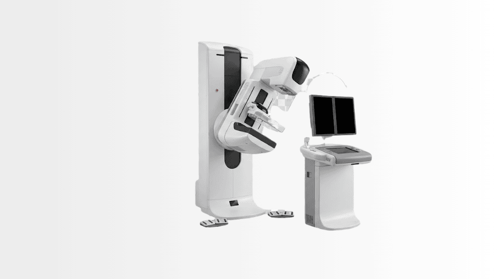
Digital mammography with 3D tomosynthesis creates layered breast images from multiple low-dose X-ray views, helping radiologists assess tissue that may be obscured on standard mammography. At Acibadem in Turkey, it is used for breast cancer screening and diagnostic evaluation when clinically appropriate.
What is a 3D mammogram (tomosynthesis)?
During the examination the machine takes a series of low-dose exposures across an arc and reconstructs them into slices about a millimetre thick. The radiologist reads the breast the way one reads a book — page by page — while a synthesised 2D image preserves the classic overview. For you, the examination feels essentially identical to a standard mammogram: the same positioning, the same brief compression, a few seconds longer per view.
Why 3D beats 2D where it matters
Large screening programmes have shown tomosynthesis finds more invasive cancers than 2D mammography alone and reduces recalls — the anxious return visits for superimposed-tissue shadows. The advantage concentrates exactly where 2D struggles most: dense breasts, where normal fibroglandular tissue is white, cancers are white, and a flat image lets one hide behind the other. Slicing separates them. Density itself matters enough that a good report states your category and what it implies for supplementary imaging.
Dense breasts: the honest conversation
Roughly half of screening-age women have dense breast tissue. Density lowers mammography’s sensitivity — 3D less so than 2D, but the effect remains — and independently raises risk modestly. The honest pathway: know your density from the report; in dense breasts, supplementary breast ultrasound can add detection, and in high-risk profiles breast MRI enters the conversation. No single tool is the whole answer; the combination is chosen with your physician against your risk profile.
The examination: what to expect
Positioning and compression mirror a standard mammogram — compression lasts seconds per view and is firm rather than painful for most women; scheduling after menstruation reduces tenderness. The radiation dose of modern tomosynthesis with synthesised 2D sits in the same low range as digital mammography — a dose regulators accept for population screening precisely because the benefit outweighs it. Results follow as a structured report with your density category and a clear recommendation.
Screening and diagnosis — both jobs
Tomosynthesis serves twice: as a screening examination on the schedule your physician recommends by age and risk, and as a diagnostic tool when something needs a closer look — a lump, discharge, a screening finding. In diagnostic work the slices localise a finding precisely enough to guide the next step, whether targeted ultrasound, MRI or a needle biopsy under imaging guidance. Within Acibadem’s breast health units, that whole pathway — imaging, biopsy, pathology, and tumour-board planning where needed — runs under one roof.
3D mammography at Acibadem
Tomosynthesis operates across the group’s breast imaging practice — with corporate-verified 3D tomosynthesis installations from Istanbul’s breast centres to City Clinic Mladost in Sofia and the Bel Medic centres in Belgrade — read by radiologists working beside the breast health teams. Comparisons with prior mammograms are part of every reading, so bring or send earlier images; they sharpen the answer.
From image to answer: how findings are graded
Every mammogram report speaks one international language: BI-RADS, the category system that turns an image into a decision. Categories 1 and 2 mean normal or definitely benign — routine screening continues. Category 3 means probably benign: a finding with a very low probability of malignancy, watched at a short interval rather than biopsied, which is a designed feature of the system rather than hedging. Categories 4 and 5 mean a biopsy should settle it — and “biopsy” today means an image-guided needle sample under local anaesthesia, not surgery: stereotactic or tomosynthesis-guided for calcifications, ultrasound-guided for masses, with results in days. Category 0 simply means more imaging is needed to decide — the recall that 3D has made rarer. Knowing the code demystifies the letter that arrives after screening: most letters are category 1–2, and even category 4 findings prove benign more often than not. The system’s whole point is that nothing suspicious waits, and nothing innocent gets over-treated.
A screening life: putting the pieces together
Breast screening is a decades-long relationship, and tomosynthesis is its workhorse rather than its entirety. The pattern that serves most women: know your personal risk once — family history, genetics where relevant, density from your first mammogram — then run the schedule your physician sets, on the same platform where possible so comparisons stay sharp. Dense-breast findings may add same-day ultrasound; high-risk profiles may add MRI on an alternating rhythm; symptoms between rounds — a new lump, skin change, discharge — always outrank the calendar and earn a diagnostic visit now, not at the next slot. Within Acibadem’s breast health units, screening, diagnostic work-up, needle biopsy, pathology and — where ever needed — tumour-board planning live on one corridor, so the distance from “the letter” to “the answer” is measured in days.
Where it is installed
18 in total Acibadem Maslak Hospital
İstanbul · 282 doctors
Acibadem Maslak Hospital
İstanbul · 282 doctors
 Acibadem Altunizade Hospital
İstanbul · 301 doctors
Acibadem Altunizade Hospital
İstanbul · 301 doctors
 Acibadem Atasehir Hospital
İstanbul · 221 doctors
Acibadem Atasehir Hospital
İstanbul · 221 doctors
 Acibadem Atakent Hospital
İstanbul · 160 doctors
Acibadem Atakent Hospital
İstanbul · 160 doctors
 Acibadem Kent Hospital (Izmir)
İzmir · 164 doctors
Acibadem Kent Hospital (Izmir)
İzmir · 164 doctors
 Acibadem Taksim Hospital
İstanbul · 64 doctors
Acibadem Taksim Hospital
İstanbul · 64 doctors
 Acibadem Adana Hospital
Adana · 109 doctors
Acibadem Adana Hospital
Adana · 109 doctors
 Acibadem Ankara Hospital
Ankara · 74 doctors
Acibadem Ankara Hospital
Ankara · 74 doctors
 Acibadem Bakirkoy Hospital
İstanbul · 110 doctors
Acibadem Bakirkoy Hospital
İstanbul · 110 doctors
 Acibadem Bodrum Hospital
Muğla · 93 doctors
Acibadem Bodrum Hospital
Muğla · 93 doctors
 Acibadem Bursa Hospital
Bursa · 106 doctors
Acibadem Bursa Hospital
Bursa · 106 doctors
 Acibadem Dr. Sinasi Can (Kadikoy) Hospital
İstanbul · 109 doctors
Acibadem Dr. Sinasi Can (Kadikoy) Hospital
İstanbul · 109 doctors
Frequently asked questions
What does my BI-RADS category mean?
It is the standard code turning your mammogram into a decision — 1–2 normal/benign, 3 short-interval watch, 4–5 biopsy to settle, 0 more imaging needed; your report states yours and the next step plainly.
If I need a biopsy, is that surgery?
No — modern breast biopsy is an image-guided needle sample under local anaesthesia, done in the imaging suite with results in days.
What if I notice a lump between screenings?
Symptoms outrank schedules — a new lump, skin change or discharge earns a diagnostic appointment now, whatever the calendar says.
Is a 3D mammogram different to have than a normal one?
Barely — same positioning, same brief compression, a few seconds longer per view while the tube sweeps its arc.
Does a 3D mammogram hurt?
Compression is firm for a few seconds per view; most women find it uncomfortable rather than painful, and scheduling after menstruation reduces tenderness.
Is the radiation higher than a standard mammogram?
Modern tomosynthesis with a synthesised 2D image sits in the same low dose range as digital mammography — accepted for routine screening because the benefit outweighs it.
Why does 3D find more cancers?
Because slicing removes overlap — the layer-by-layer read separates a real mass from stacked normal tissue, exactly where flat images struggle.
Why are fewer women called back after 3D screening?
Most recalls after 2D screening are superimposition shadows; slices dissolve them at the first reading instead of at a second appointment.
What does breast density mean for me?
Dense tissue lowers mammographic sensitivity and modestly raises risk — your report states your category, and in dense breasts supplementary ultrasound or, in high-risk profiles, MRI may be advised.
At what age should mammography screening start?
Recommendations vary by risk profile and country; your physician sets your starting age and interval against your personal and family history.
How can we help you today?
Send your diagnosis and reports — a senior specialist reviews them and replies.
Send my reportsMeet an Acıbadem specialist by secure video, before you travel.
Book a video visit